Anti-Mouse IgG DL680, TrueBlot, DL680 TrueBlot ULTRA, DyLight(TM) 680 TrueBlot, TrueBlot for IP/WB, TrueBlot for immunoprecipitation, TrueBlot for western blotting, Fluorescent TrueBlot, Ms TrueBlot, IRDye 700, IRDye 680
Clonality:
Monoclonal
Concentration:
1.0 mg/mL by UV absorbance at 280 nm
Clone Designation:
[eB144]
Buffer:
0.02 M Potassium Phosphate, 0.15 M Sodium Chloride, pH 7.2
Form:
Lyophilized
Target:
Mouse
Application Dilute:
FLISA: User Optimized, Flow Cytometry: 1:2,000 - 1:10,000, IHC: User Optimized, IF Microscopy: 1:500 - 1:2,500, WB: 1:1000
Application Notes:
Fluorescent Mouse TrueBlot Antibody DyLight(TM) 680 has been tested in ELISA, immunofluorescence, immunoprecipitation, and western blot and may also be used for detection in immunoassays that do not employ immunoprecipitation. Fluorescent Mouse TrueBlot A
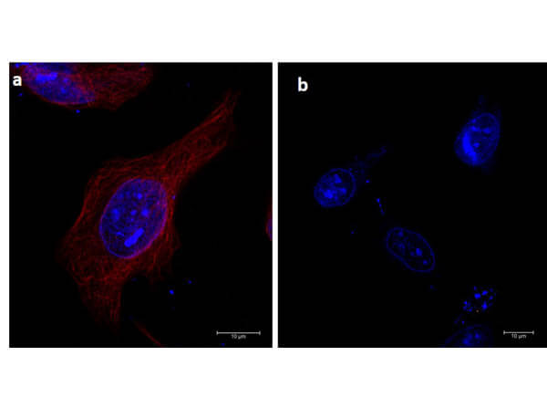
Immunofluorescence microscopy of alpha-tubulin in HeLa cells using DyLight(TM) 680-conjugated Fluorescent TrueBlot anti-mouse IgG (p/n 18-4417-32) for detection. HeLa cells were fixed with 100% methanol, blocked (5% rat serum/0.3% Triton X-100 in 1X PBS) for 1hr, then incubated with 15µg/mL of anti-alpha-tubulin primary antibody (p/n 200-301-880) at 4C overnight. Following 3 washes in 1X PBS for 5 min each, 5µg/mL of Fluorescent TrueBlot anti-mouse IgG DyLight(TM) 680 was added and allowed to incubate for 1hr at room temperature. Nuclei were counterstained with DAPI present in mounting medium. The predicted main localization is microtubules. Image taken at 63X magnification. (a) Merged a-tubulin (red)/DAPI (blue) image shown. (b) secondary antibody only.
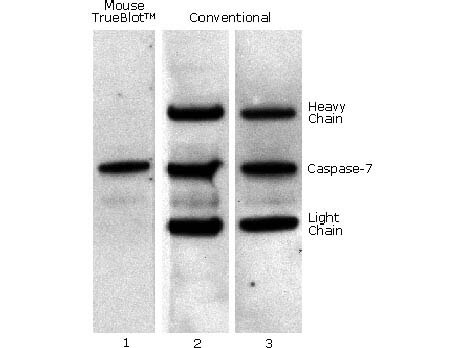
Mouse TrueBlot IP / Western Blot: Caspase 7 was immunoprecipitated from 0.5 ml of 1x10e7 Jurkat cells/ml with 5 ug mouse anti-human Caspase 7. Precipitate from 1x10e6 cells was subjected to electrophoresis, transferred to an PVDF membrane, and Western blotted with anti-Caspase 7 using Mouse TrueBlot ULTRA: Anti-Mouse Ig HRP (Lane 1) or conventional HRP-conjugated anti-mouse antibody (Lane 2) - note the detection of the heavy and light chains of the immunoprecipitating antibody in Lane 2 but not in Lane 1. When Lane 1 is re-immunoblotted using conventional HRP-conjugated anti-mouse polyclonal antibody (Lane 3), the heavy and light chains are now detected, confirming that although the immunoprecipitating heavy and light chains are present, Mouse TrueBlot ULTRA: Anti-Mouse Ig HRP detects only native antibody and not denatured heavy and light chains.
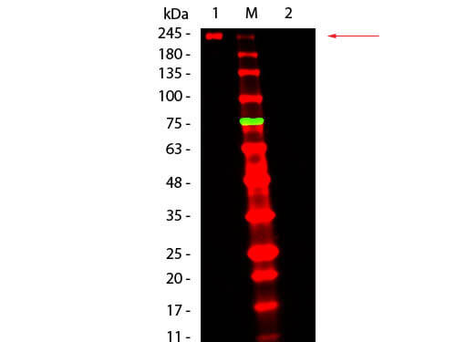
Western Blot of Fluorescent TrueBlot: Anti-Mouse Ig DyLight 680 Conjugated. Lane 1: Mouse IgG, Non-reduced. Lane 2: Mouse IgG, Reduced. Load: 50 ng per lane. Primary antibody: none. Secondary antibody: Fluorescent TrueBlot: Anti-Mouse Ig DyLight 680 Conjugated at 1:1,000 for 60 min at RT. Block: MB-070 for 30 min at RT. Predicted/Observed size: 160 kDa for Mouse IgG, Non-reduced. Migrates at slightly higher molecular weight. Other band(s): none.
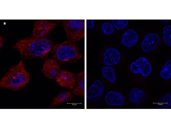
Immunofluorescence microscopy of a-tubulin in A431 cells using DyLight(TM) 680-conjugated Fluorescent TrueBlot anti-mouse IgG for detection. A431 cells were fixed with 100% methanol, blocked (5% rat serum/0.3% Triton X-100 in 1X PBS) for 1 hr, then incubated with 15 µg/mL of anti-a-tubulin primary antibody (Cat. No. 200-301-880) at 4C overnight. After 3 washes in 1X PBS for 5 min each, 5 µg/mL of Fluorescent TrueBlot anti-mouse IgG DyLight(TM) 680 was added and allowed to incubate for 1 hr at room temperature. Nuclei were counterstained with DAPI present in mounting medium. The predicted main localization is microtubules. Image taken at 63X magnification. (a) Merged a-tubulin (red)/DAPI (blue) image shown. (b) secondary antibody only.
* VAT and and shipping costs not included. Errors and price changes excepted