Anti-AKT Antibody was produced by repeated immunizations in mice with a synthetic peptides corresponding to residues internal to the human AKT1, AKT2, and AKT3 proteins.
Mouse Anti-AKT Antibody is tested for ELISA and western blotting. This antibody is suitable for immunohistochemistry and immunoprecipitation. Expect a band approximately 54 - 56 kDa in size corresponding to AKT protein by western blotting in the appropri
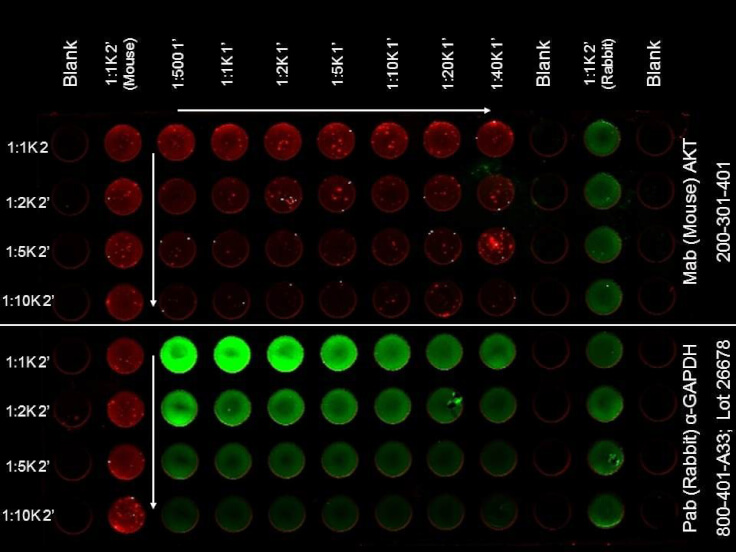
ELISA of Mouse Anti-AKT Antibody. Antigen: HCT-116 cell line (p/n W09-001-GM4). Coating amount: Confluent in the 96 well plate. Primary antibody: AKT (top) or GAPDH (bottom) antibody at 2 µg/mL. Dilution series: Primary and Secondary Antibodies 2-fold. Mid-point concentration: N/A. Secondary antibody: DyLight(TM) 680 donkey secondary antibody and DyLight(TM) 800 goat secondary antibody starting at 1:1,000. Substrate: None.
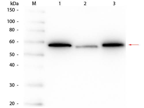
Western Blot of Mouse anti-AKT Monoclonal Antibody. Lane 1: His-AKT1 Recombinant Protein. Lane 2: His-AKT2 Recombinant Protein. Lane 3: His-AKT3 Recombinant Protein. Load: 50 ng per lane. Primary antibody: Mouse anti-AKT Monoclonal Antibody at 1:1,000 overnight at 4C. Secondary antibody: HRP mouse secondary antibody at 1:40,000 for 30 min at RT. Block: MB-070 for 30 min at RT. Predicted/Observed size: 54-56 kDa, 54-56 kDa for AKT1, AKT2, AKT3.
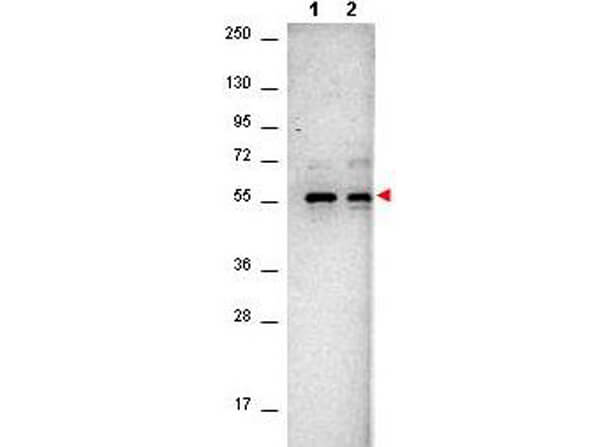
Western Blot of Mouse anti-AKT antibody. Lane 1: NIH/3T3 cell lysates (p/n W10-000-358). Lane 2: PDGF stimulated NIH/3T3 cell lysates (p/n W10-001-377). Load: 10µg per lane. Primary antibody: Anti-AKT antibody at 1:400 for overnight at 4C. Secondary antibody: Goat-anti-Mouse IgG HRP conjugated (p/n 610-103-121) was used at a 1:40,000 dilution for 1hr at 2-8C with FemtoMax(TM) enhanced chemiluminescent reagent (p/n FEMTOMAX-100). Block: 5% BLOTTO (p/n B501-0500) in TBS for 2hr at RT. Observed size: ~56 kDa for AKT.
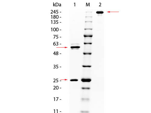
SDS PAGE of Mouse anti-AKT Monoclonal Antibody. Lane 1: Reduced Mouse anti-AKT Monoclonal Antibody. Lane M: 3 µL Opal Prestained Marker (p/n MB-210-0500). Lane 2: Non-Reduced Mouse anti-AKT Monoclonal Antibody. Load: 1 µg per lane. Predicted/Observed size: Non-Reduced at 160kDa/Observed at 245 kDa, Reduced at 55, 25 kDa. Non-reduced migrates slightly higher.
* VAT and and shipping costs not included. Errors and price changes excepted