Hemagglutinin of A/Vietnam/1203/04 Influenza Virus (VN04-8) monoclonal antibody was produced by intraperitoneal immunization of BALB/c mice with concentrated purified virus preparation containing hemagglutinin (HA) protein of influenza A virus [strain A/Vietnam/1203/04 (H5N1)] using the modification of the method described by Kohler and Milstein. Each mouse received two immunizations of 15 µg HA with incomplete Freunds adjuvant, administered 3 week apart.
0.02 M Potassium Phosphate, 0.15 M Sodium Chloride, pH 7.2
Form:
Liquid (sterile filtered)
Target:
H5N1 virus
Antibody Type:
Primary Antibody
Application Dilute:
ELISA: 1:5,000, IHC: User Optimized, IP: User Optimized, WB: User Optimized
Application Notes:
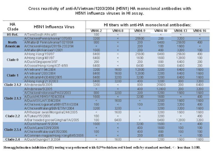
Hemagglutinin of A/Vietnam/1203/04 Influenza Virus (VN04-8) monoclonal antibody can be used for hemagglutination inhibition (HI) assays to provide antigenic characterization of the influenza A viruses of the H5 HA subtype. This monoclonal antibody is sui
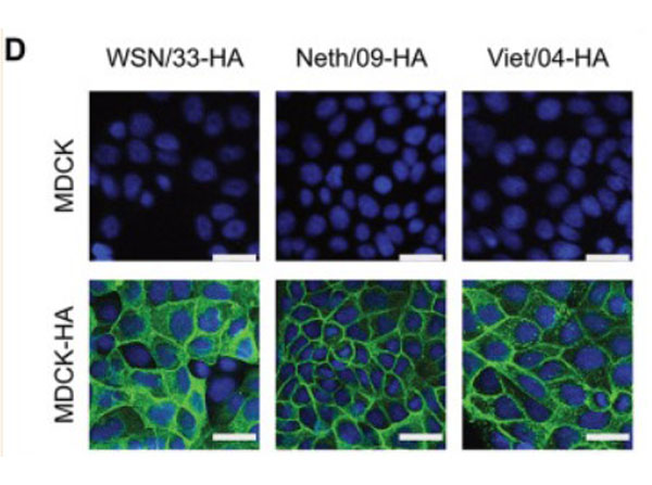
Immunofluorescence of Anti-H5N1 Antibody.Biologically contained WSN/33, 2009 pandemic H1N1, and highly pathogenic H5N1 IAVs expressingRenillaluciferase. (D) Indirect immunofluorescence analysis of MDCK and MDCK-HA cell lines stably expressing each strain-specific HA (green). DAPI was used to stain cell nuclei (blue). Bars, 25µm. Fig 1. PMID: 32848003.
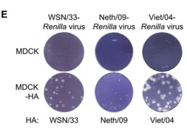
Biologically contained WSN/33, 2009 pandemic H1N1, and highly pathogenic H5N1 IAVs expressingRenillaluciferase. (E) Plaque formation assay for each IAV reporter on MDCK and MDCK-HA cell lines stably expressing each strain-specific HA. Each cell line was infected with the same number of PFU, and assays were fixed and stained 36 h later. Fig 1. PMID: 32848003.
Western Blot of Anti-H5N1 Antibody. Biologically contained WSN/33, 2009 pandemic H1N1, and highly pathogenic H5N1 IAVs expressingRenillaluciferase. (C) Western blot analysis of MDCK and MDCK-HA cell lines stably expressing each strain-specific HA. Specific antibodies were used to detect HA and actin. Fig 1. PMID: 32848003.
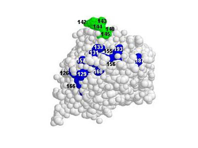
Schematic representation of the antigenic sites and the epitopes on the globular head of the HA H5 HA molecule. Images were created with RasMol 2.6, and the HA structure was obtained from the Protein Data Bank (PDB accession number 1JSM). Amino acid positions are designated in H3 numbering. Image provided courtesy of Elena Govorkova Ph D.
* VAT and and shipping costs not included. Errors and price changes excepted