Anti-AKT1 Antibody was produced in mice by repeated immunizations with a synthetic peptide corresponding to internal residues of human AKT1 protein followed by monoclonal development.
0.02 M Potassium Phosphate, 0.5 M Sodium Chloride, pH 7.2
Form:
Lyophilized
Target:
Human
Antibody Type:
Primary Antibody
Application Dilute:
ELISA: User Optimized, Flow Cytometry: User Optimized, IHC: User Optimized, IF Microscopy: User Optimized, WB: User Optimized
Application Notes:
Anti-AKT1 BIOTIN Antibody is suitable for Flow Cytometry, ELISA, immunohistochemistry, and western blotting. Expect a band approximately 56 kDa in size corresponding to AKT1 protein by western blotting in the appropriate cell lysate or extract. This mono
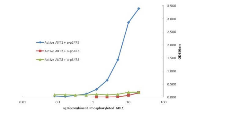
Plate was coated with monoclonal anti AKT1 antibody (capture antibody) followed by incubation with recombinant AKT1 (p/n 009-001-P21), AKT2 (p/n 009-001-P22), AKT3 (p/n 009-001-P23) proteins. Binding was detected with biotinylated monoclonal anti-AKT pS473. The signal shows specificity of the monoclonal anti-AKT1 antibody to recombinant isoform AKT1 protein and not the isoform 2 and 3.
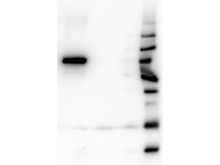
Western Blot of Mouse Anti-AKT1 antibody. Lane 1: GST Tagged recombinant AKT1. Lane 2: GST Tagged recombinant AKT2. Lane 3: GST Tagged recombinant AKT3. Load: 25 ng per lane. Primary antibody: AKT1 antibody at 1:1,000 for overnight at 4C. Secondary antibody: Peroxidase mouse secondary antibody at 1:40,000 for 30 min at RT. Block: MB-070 for 30 min at RT. Predicted/Observed size: 78 kDa for AKT1. Other band(s): none.
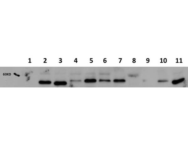
Western Blot of Mouse Anti-AKT1 antibody. Lane 1: AKT-1 Null. Lane 2: WT. Lane 3: MEF 1. Lane 4 : A549 (p/n W09-001-GX4). Lane 5: Calu-1. Lane 6: PC-3 (p/n W09-001-GV6). Lane 7: HepG2 (p/n W09-001-GJ5). Lane 8: Jurkat (p/n W09-001-370). Lane 9: SKOV3 (p/n W09-001-GX9). Lane 10: HEK293T (p/n W09-001-GX5). Lane 11: C2C12 (p/n W10-001-GL7). Load: 20 ug per lane. Primary antibody: AKT1 antibody at 1:1,000 for overnight at 4C. Secondary antibody: Peroxidase mouse secondary antibody at 1:40,000 for 30 min at RT. Block: MB-070 for 30 min at RT. Predicted/Observed size: 56 kDa for AKT1. Other band(s): none.
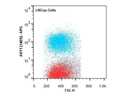
Flow Cytometry of Mouse anti-AKT1 antibody. Cells: LNCap Cells. Stimulation: none. Primary antibody: Allophycocyanin AKT1 antibody at 1.0 µg/mL for 20 min at 4C.
* VAT and and shipping costs not included. Errors and price changes excepted