NANOG Antibody was produced from whole rabbit serum prepared by repeated immunizations with a peptide corresponding to an internal region of human NANOG.
Conjugation:
Unconjugated
Alternative Names:
NANOG homeobox, Homeobox transcription factor Nanog, hNanog, Homeobox protein NANOG
0.02 M Potassium Phosphate, 0.15 M Sodium Chloride, pH 7.2
Form:
Liquid (sterile filtered)
Target:
Human
Antibody Type:
Primary Antibody
Application Dilute:
IHC: 5µg/mL, IF Microscopy: 20µg/mL, WB: 1-2 ug/mL
Application Notes:
Anti-NANOG Antibody is tested for use in E, WB, IF, and IHC. Expect a band approximately ~34.6 kDa on specific lysates. Western Blot, Immunohistochemistry, and Immunofluorescence tested in human samples. Specific conditions for reactivity should be optim
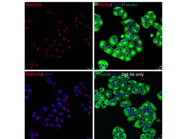
Immunofluorescence Validation of NANOG. Cell: HeLa cells. Fixation: PFA-fixed. Primary Antibody: Anti-NANOG at 20 µg/mL. Secondary: goat anti-rabbit IgG secondary antibody at 1:1000 dilution (red) and DAPI staining (blue). Alpha tubulin was stained with anti-alpha tubulin antibody following by goat anti-mouse IgG secondary antibody (green). Images were captured with confocal microscopy.
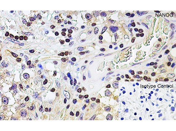
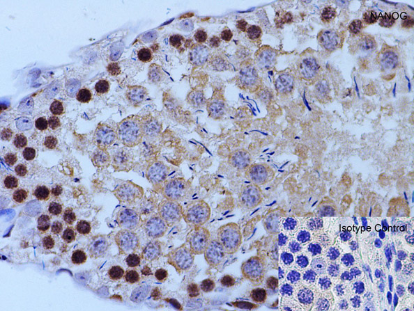
Immunohistochemistry of NANOG. Tissue: human testis tissue. Fixation: paraffin-embedded, formaldehyde and blocked with 10% serum for 1 h at RT. Antigen retrieval: heat mediation with a citrate buffer (pH6). Primary Antibody: anti-NANOG antibody at 5 µg/ml overnight at 4C. Secondary: goat anti-rabbit IgG H&L (HRP) at 1:250. Counter stained with Hematoxylin.
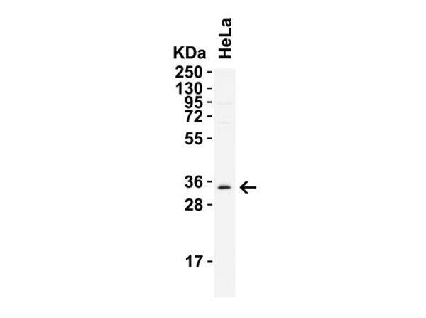
Western Blot of NANOG. Load: 15 µg of HeLa cell lysate. Primary antibody: Anti-NANOG at 2 µg/mL for 1h incubation at RT in 8% NFDM/TBST. Secondary: Goat Anti-Rabbit IgG HRP conjugate at 1:10,000 dilution.
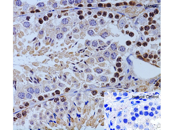
Immunohistochemistry Validation of NANOG. Tissue: mouse testis tissue. Fixation: paraffin-embedded, formaldehyde and blocked with 10% serum for 1 h at RT. Antigen retrieval: heat mediation with a citrate buffer (pH6). Primary Antibody: anti-NANOG antibody at 2 µg/ml overnight at 4C. Secondary: goat anti-rabbit IgG H&L (HRP) at 1:250. Counter stained with Hematoxylin.
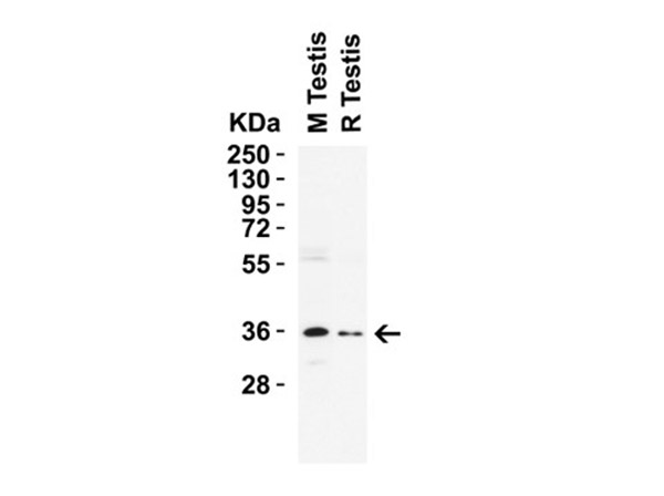
Western Blot of NANOG. Load: 15 µg of lysates per lane. Lane 1: mouse testis, Lane 2 : rat testis. Primary antibody: Anti-NANOG at 2 µg/mL for 1h incubation at RT in 8% NFDM/TBST. Secondary: Goat anti-rabbit IgG HRP conjugate at 1:10,000 dilution.
Immunohistochemistry Validation of NANOG. Tissue: rat testis tissue. Fixation: paraffin-embedded, formaldehyde and blocked with 10% serum for 1 h at RT. Antigen retrieval: heat mediation with a citrate buffer (pH6). Primary Antibody: anti-NANOG antibody at 2 µg/ml overnight at 4C. Secondary: goat anti-rabbit IgG H&L (HRP) at 1:250. Counter stained with Hematoxylin.
* VAT and and shipping costs not included. Errors and price changes excepted