Anti-Connexin 43 antibody was prepared from whole rabbit serum produced by repeated immunizations with a synthetic peptide corresponding to an internal portion of mouse connexin 43 conjugated to Keyhole Limpet Hemocyanin (KLH).
Conjugation:
Unconjugated
Alternative Names:
Rabbit Anti-Connexin 43 Antibody, Rabbit Anti-Gja1 Antibody, Gap Junction Protein Alpha 1, Gap Junction Protein, Alpha 1, 43kDa, Gap Junction 43 KDa Heart Protein, Connexin-43, GJAL, Gap Junction Protein, Alpha 1, 43kDa (Connexin 43), Oculodentodigital Dysplasia (Syndactyly Type III), Gap Junction Protein, Alpha-Like, Gap Junction Alpha-1 Protein, Connexin 43, AVSD3, EKVP3, HLHS1, PPKCA, CMDR, CX43, EKVP, ODDD, Cx43, HSS
Anti-Connexin 43 Antibody has been tested in ELISA, WB, IF, and IHC. Expect a band at ~43Da in western blot using appropriate lysates. Positive control used: Mouse Heart in IHC, Rat Pup Brain cells, NIH-3T3, or HEK293T in IF, and HEK293T, Mouse Heart, or
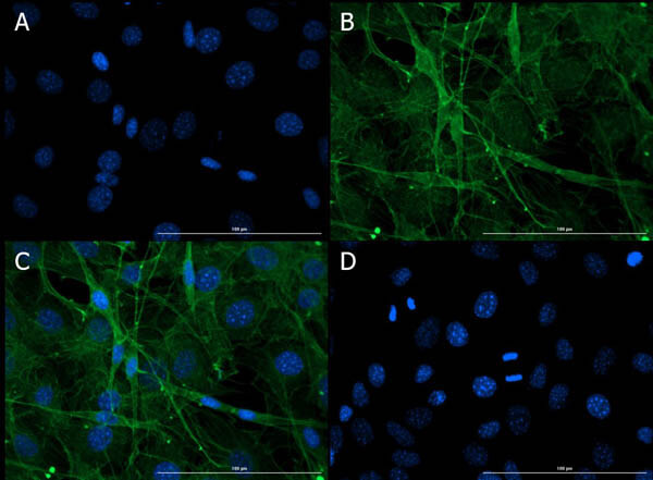
Immunofluorescence of Rabbit Anti-Connexin 43 Antibody. Cell Line: NIH-3T3 Cells. Fixative: 4% PFA. Permeabilization: 0.3% Triton X-100. Primary Antibody: Anti-Connexin 43 Antibody at 15µg/mL overnight at 2-8C. Secondary Antibody: Donkey Anti-Rabbit IgG DyLight(TM)488 (p/n 611-741-127) at 5µg/mL for 1hr at RT. Nuclear Counterstain: DAPI. Staining: (A). DAPI. (B). Primary + Secondary Antibody. (C). Merged A+B. (D). Secondary Only. Expected localization: cell membrane, ER - (UniProtID). Vesicles, cell junctions, nucleoplasm - (Human Protein Atlas).
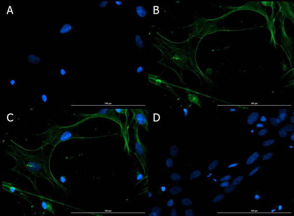
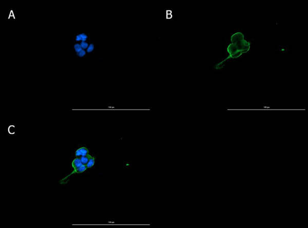
Immunofluorescence of Rabbit Anti-Connexin 43 Antibody. Cell Line: Rat Pup Brain Cells. Fixative: 4% PFA. Permeabilization: 0.3% Triton X-100. Primary Antibody: Anti-Connexin 43 Antibody at 15µg/mL overnight at 2-8C. Secondary Antibody: Donkey Anti-Rabbit IgG DyLight(TM)488 (p/n 611-741-127) at 5µg/mL for 1hr at RT. Nuclear Counterstain: DAPI. Staining: (A). DAPI. (B). Primary + Secondary Antibody. (C). Merged A+B. (D). Secondary Only. Expected localization: cell membrane, ER - (UniProtID). Vesicles, cell junctions, nucleoplasm - (Human Protein Atlas).
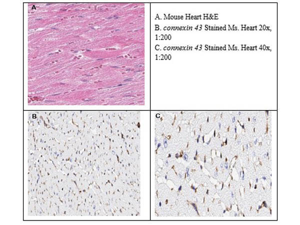
Immunohistochemistry of Rabbit Anti-Connexin 43 Antibody. Tissue: Mouse Heart. Antigen Retrieval: HIER citrate buffer for 20 min. Primary Antibody: Anti-Connexin 43 at 1:200 for 30min at RT. Secondary Antibody: Anti-Rabbit Poly IgG-HRP Ready-to-Use for 8 min at RT. Stain: DAB. Counterstain: Hematoxylin. Connexin 43 showed moderate to intense staining of myocytes and vascular endothelium.
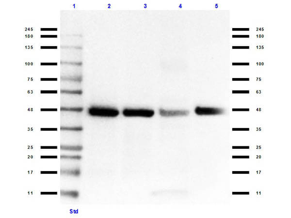
Western Blot of Rabbit Anti-Connexin 43 Antibody. Lane 1: PreStained Opal Molecular Weight Ladder (p/n MB-210-0500). Lane 2: Connexin HEK293T overexpressing lysate (10µg). Lane 3: HEK293T lysate (p/n W09-001-GX5) [10µg]. Lane 4: Mouse Heart lysate (p/n W10-000-T014) [35µg]. Lane 5: Rat Brain lysate (p/n W12-000-T077) [35µg]. Primary Antibody: Anti-Connexin 43 Antibody at 1:1000 overnight at 2-8C. Secondary Antibody: Goat Anti-Rabbit HRP (p/n 611-103-122) at 1:70,000 for 30min at RT. Buffer: BlockOut (p/n MB-073). Predicted: ~43kDa for ms, rt, hu.
Immunofluorescence of Rabbit Anti-Connexin 43 Antibody. Cell Line: HEK293T Cells. Fixative: 4% PFA. Permeabilization: 0.3% Triton X-100. Primary Antibody: Anti-Connexin 43 Antibody at 15µg/mL overnight at 2-8C. Secondary Antibody: Donkey Anti-Rabbit IgG DyLight(TM)488 (p/n 611-741-127) at 5µg/mL for 1hr at RT. Nuclear Counterstain: DAPI. Staining: (A). DAPI. (B). Primary + Secondary Antibody. (C). Merged A+B. Expected localization: cell membrane, ER - (UniProtID). Vesicles, cell junctions, nucleoplasm - (Human Protein Atlas).
* VAT and and shipping costs not included. Errors and price changes excepted