Anti-Smad1 antibody was prepared from whole rabbit serum produced by repeated immunizations with a synthetic peptide corresponding to an internal portion of mouse Smad1 conjugated to Keyhole Limpet Hemocyanin (KLH).
Conjugation:
Unconjugated
Alternative Names:
Mothers against decapentaplegic homolog 1, MAD homolog 1, Mothers against DPP homolog 1, Dwarfin-A, Dwf-A, Mothers-against-DPP-related 1, Mad-related protein 1, mMad1, SMAD 1, Smad1, Madh1, Madr1
0.02 M Potassium Phosphate, 0.15 M Sodium Chloride, pH 7.2
Form:
Liquid (sterile filtered)
Target:
Mouse
Antibody Type:
Primary Antibody
Application Dilute:
ELISA: 5µg/ml, IHC: 1:400-1:500, IF Microscopy: 5-10µg/mL, WB: 5µg/mL
Application Notes:
Anti-Smad1 Antibody has been tested in ELISA, WB, IF, and IHC. Expect a band at ~52.2kDa in western blot using appropriate tissues or lysates. Positive control used: Mouse testis and C2C12 in Western Blot, C2C12 in Immunofluorescence, Mouse Lung, Skeleta
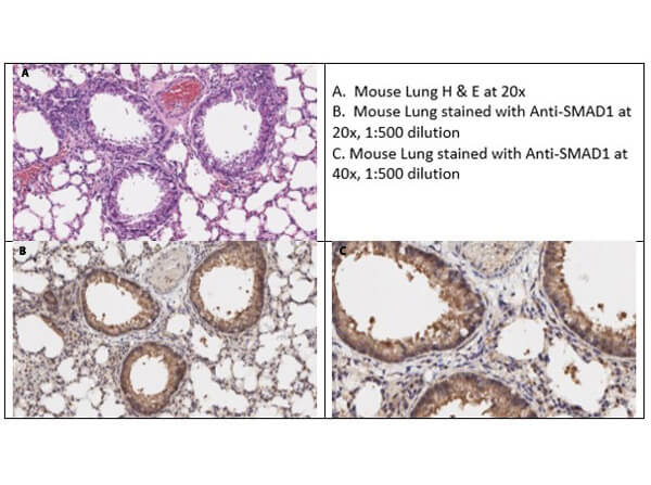
Immunohistochemistry of Rabbit Anti-Smad1 Antibody. Tissue: Mouse Lung Tissue. Fixative: none. Antigen Retrieval: HIER using citrate buffer for 20mins. Primary Antibody: Anti-Smad1 at 1:500 for 30mins at RT. Secondary Antibody: Anti-Rabbit poly HRP IgG Ready-to-Use for 8mins at RT. Counterstain: Hematoxylin. Substrate: DAB. Analysis Report: Smad1 shows strong cytoplasmic staining of airway bronchial epithelium and of mesenchymal cells within the alveolar walls in mouse lung tissue at 1:500.
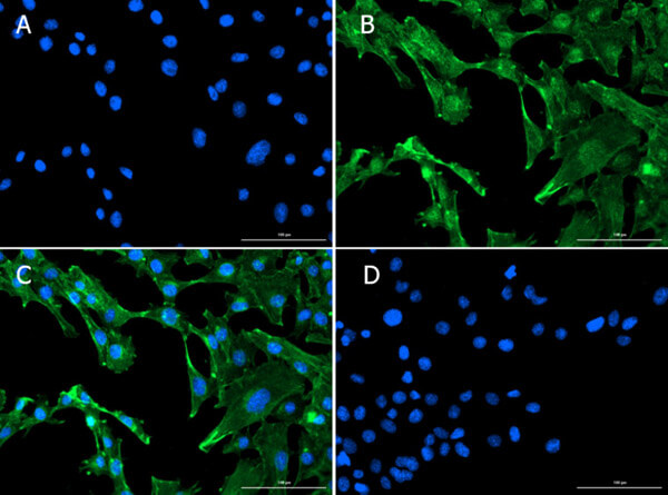
Immunofluorescence of Rabbit Anti-Smad1 Antibody. Cells: C2C12 cells. Fixative: 100% MeOH. Permeabilization: 0.3% Triton X-100. Primary Antibody: Anti-Smad1 at 10µg/mL overnight at 2-8C. Secondary Antibody: Donkey Anti-Rabbit IgG DyLight(TM)488 (p/n 611-741-127) at 5µg/mL for 1hr at RT. Nuclear Counterstain: DAPI. Staining: (A) DAPI. (B) Anti-Smad1 + DyLight(TM)488. (C) Merge A+B. (D) Secondary Only. Expected localization: Nucleus, Plasma membrane, andCytosol.
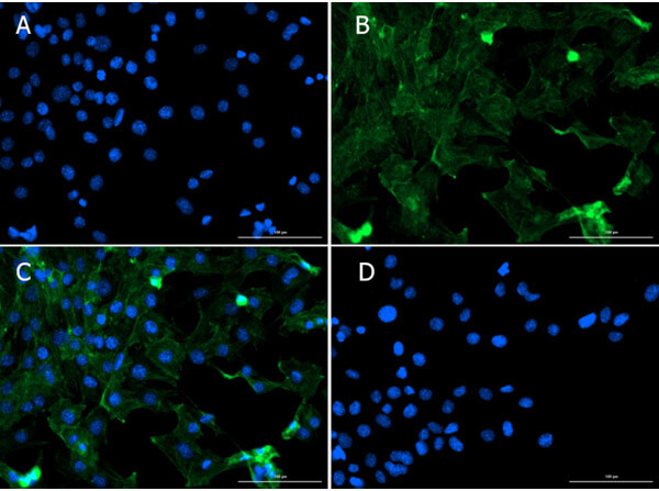
Immunofluorescence of Rabbit Anti-Smad1 Antibody. Cells: C2C12 cells. Fixative: 100% MeOH. Permeabilization: 0.3% Triton X-100. Primary Antibody: Anti-Smad1 at 5µg/mL overnight at 2-8C. Secondary Antibody: Donkey Anti-Rabbit IgG DyLight(TM)488 (p/n 611-741-127) at 5µg/mL for 1hr at RT. Nuclear Counterstain: DAPI. Staining: (A) DAPI. (B) Anti-Smad1 + DyLight(TM)488. (C) Merge A+B. (D) Secondary Only. Expected localization: Nucleus, Plasma membrane, and Cytosol.
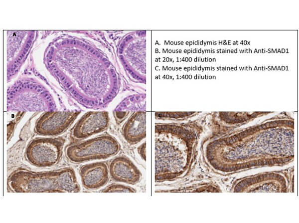
Immunohistochemistry of Rabbit Anti-Smad1 Antibody. Tissue: Mouse Testis. Fixative: none. Antigen Retrieval: HIER using citrate buffer for 20mins. Primary Antibody: Anti-Smad1 at 1:400 for 30mins at RT. Secondary Antibody: Anti-Rabbit poly HRP IgG Ready-to-Use for 8mins at RT. Counterstain: Hematoxylin. Substrate: DAB. Analysis Report: Membranous/cytoplasmic staining is seen in the epithelial cells lining the epididymis.
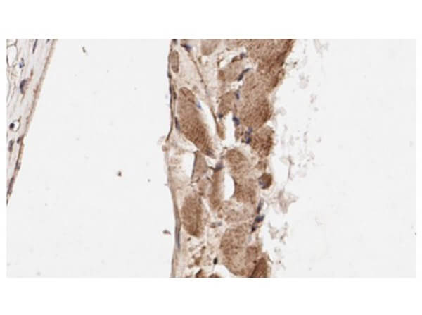
Immunohistochemistry of Rabbit Anti-Smad1 Antibody. Tissue: Mouse Skeletal Muscle Tissue. Fixative: none. Antigen Retrieval: HIER using citrate buffer for 20mins. Primary Antibody: Anti-Smad1 at 1:400 for 30mins at RT. Secondary Antibody: Anti-Rabbit poly HRP IgG Ready-to-Use for 8mins at RT. Counterstain: Hematoxylin. Substrate: DAB. Analysis Report: Cytoplasmic staining of skeletal muscle myocytes is seen.
* VAT and and shipping costs not included. Errors and price changes excepted