0.01 M Sodium Phosphate, 0.25 M Sodium Chloride, pH 7.2
Form:
Liquid (sterile filtered)
Target:
Human
Antibody Type:
Primary Antibody
Application Dilute:
ELISA: 1:1,000, ChIP: 2 µg/ChIP, IF Microscopy: 1:500, WB: 1:1,000
Application Notes:
Anti-H3K27me2 Antibody is tested in Chromatin Immunoprecipitation, Dot Blot, ELISA, Immunofluorescence, and Western Blots. Specific conditions for reactivity should be optimized by the end user. Expect a band approximately 17 kDa in the appropriate cell
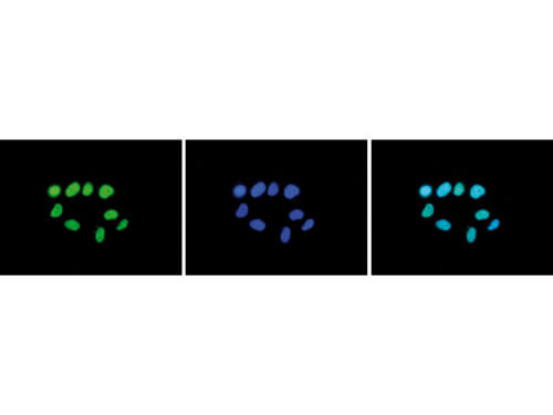
Immunofluorescence Microscopy results of Rabbit anti-Histone H3 K27 me2 antibody. Tissue: HeLa cells. Fixation: 4% formaldehyde for 10 min. Block: PBS/Triton X-100 / 5% normal goat serum and 1% BSA. Primary antibody: Histone H3 K27 me2 antibody at 1:500 for 1 hr at RT. Secondary antibody: anti-rabbit Alexa 488 secondary antibody at 1:10,000 for 45 min at RT. Staining: Histone H3 K27 me2 antibody as green fluorescent signal (left), with DAPI (middle), and a merge of the two stainings (right).
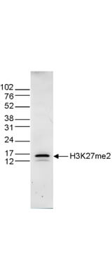
Western Blot results of Rabbit anti-Histone H3 K27 me2 antibody. Lane 1: 15 µg of histone extracts from HeLa cells. Primary antibody: Rabbit anti-Histone H3 K27 me2 antibody at 1:1,000 overnight at 4C. Secondary antibody: Peroxidase anti-rabbit secondary antibody at 1:10,000 for 45 min at RT. Block: TBS-Tween / 5% BLOTTO. Predicted/Observed size: ~15 kDa for Rabbit Histone H3 K27 me2.
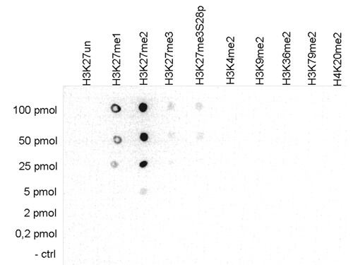
Dot Blot results of Rabbit anti-Histone H3 K27 me2 antibody. Antigens: H3K27, H3K27me1, H3K27me2, H3K27me3, H3K27me3 S28p, H3K4me2, H3K9me2, H3K36me2, H3K79me2, H4K20me2. Load: 100pmol, 50pmol, 25pmol, 5pmol, 2pmol, 0.2pmol, and control. Primary antibody: Histone H3 K27 me2 antibody at 1:50,000 for 45 min at 4C. Secondary antibody: anti-rabbit HRP secondary antibody at 1:10,000 for 45 min at RT. Block: 5% BLOTTO.
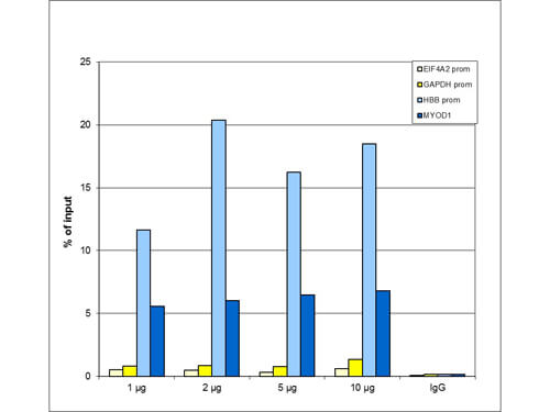
Chromatin Immunoprecipitation Rabbit H3K27me2 Antibody. ChIP assays were performed using HeLa cells, the Anti-H3K27me2 Antibody, and optimized PCR primer sets for qPCR. ChIP was performed using sheared chromatin from 1 million cells. A titration of the antibody consisting of 1, 2, 5, and 10 µg per ChIP experiment was analyzed. IgG (2 µg/IP) was used as negative IP control. QPCR was performed with primers for the promoter of the active GAPDH and EIF4A2 genes, used as negative controls, and for the promoter of the inactive HBB and the coding region of the inactive MYOD1 genes, used as positive controls. This figure shows the recovery, expressed as a % of input (the relative amount of immunoprecipitated DNA compared to input DNA after qPCR analysis). These results are in accordance with the observation that H3K27me2 is preferably present at silent genes.
* VAT and and shipping costs not included. Errors and price changes excepted