Anti-Histone H3 K27 me1 Antibody was produced in rabbits by repeated immunizations with a synthetic peptide of histone H3 containing the monomethylated lysine 27.
0.01 M Sodium Phosphate, 0.25 M Sodium Chloride, pH 7.2
Form:
Liquid (sterile filtered)
Target:
Human
Antibody Type:
Primary Antibody
Application Dilute:
ELISA: 1:500, ChIP: 1 µg per IP, IF Microscopy: 1:1,000, WB: 1:1,000
Application Notes:
Anti-Histone H3 K27 me1 Antibody is tested in Chromatin Immunoprecipitation, Dot Blot, ELISA, Immunofluorescence, and Western Blots. Specific conditions for reactivity should be optimized by the end user. Expect a band approximately 15kDa in the appropri
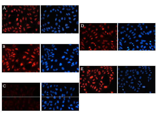
Immunofluorescence results of Anti-Histone H3K27me1 Antibody. Tissue: Human osteosarcoma (U2OS) cells. Fixation: 4% formaldehyde for 20 min. Block: PBS/Triton X-100 / 5% normal goat serum. Incubation: Figure A-E: H3K27me1 antibody 1:1,000. Figure B: 2ng/µl H3K27 peptide. Figure C: 2ng/µl H3K27me1 peptide. Figure D: 2ng/µl H3K27me2 peptide. Figure E: 2ng/µl H3K27me3 peptide. Secondary antibody: anti-rabbit Alexa568 secondary antibody at 1:10,000 for 45 min at RT (Left) or DAPI (right). Staining: Histone H3K27me1 antibody as (red) fluorescent signal, DAPI (blue).
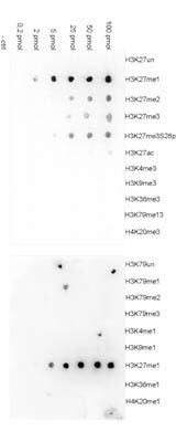
Dot Blot results of H3K27me1 antibody. Antigens: H3K27me1 and other modifications and unmodified sequences of histone H3 and H4. Load: 100 pmol, 50 pmol, 25 pmol, 5 pmol, 2 pmol, and 0.2 pmol with control. Primary antibody: H3K27me1 antibody at 1:20,000 for 45 min at 4C. Secondary antibody: anti-rabbit HRP antibody at 1:20,000 for 45 min at RT. Block: 5% BLOTTO.
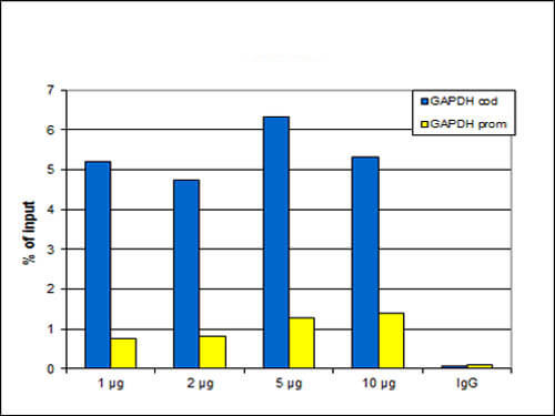
Chromatin Immunoprecipitation Rabbit Anti-H3K27me1 Antibody. ChIP assays were performed using HeLa cells, H3K27me1 Antibody, and optimized PCR primer sets for qPCR. ChIP was performed using sheared chromatin from 100,000 cells. A titration of the antibody consisting of 1, 2, 5 and 10 µg per ChIP experiment was analyzed. IgG (2 µg/IP) was used as negative IP control. qPCR was performed with primers for the promoter and the coding region of the active gene GAPDH used as a negative and a positive control target, respectively. This figure shows the recovery, expressed as a % of input (the relative amount of immunoprecipitated DNA compared to input DNA after qPCR analysis).
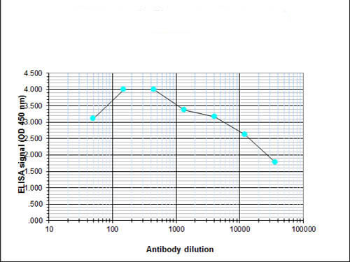
ELISA results of H3K27me1 antibody. Antigen: BSA coupled to H3K27me1. Coating amount: 0.1 µg per well. Dilution series: serial. Titer: 1:32,900 H3K27me1 antibody. Substrate: TMB (p/n TMBE-1000).
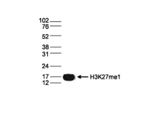
Western Blot results of Rabbit anti-Histone H3K27me1 antibody. Lane 1: 15 µg of histone extracts from HeLa cells. Primary antibody: Rabbit anti-Histone H3 K27 me2 antibody at 1:1,000. Secondary antibody: Peroxidase anti-rabbit secondary antibody at 1:10,000 for 45 min at RT. Block: TBS-Tween / 5% BLOTTO. Predicted/Observed size: ~15 kDa for Rabbit Histone H3 K27 me2.
* VAT and and shipping costs not included. Errors and price changes excepted