0.02 M Potassium Phosphate, 0.15 M Sodium Chloride, pH 7.2
Form:
Lyophilized
Target:
Goat
Antibody Type:
Secondary Antibody
Application Dilute:
FLISA: >1:20,000, IF Microscopy: >1:5,000, WB: >1:10,000
Application Notes:
Anti-Goat IgG DyLight 488 Antibody has been tested by dot blot. This product is designed for immunofluorescence microscopy, fluorescence based plate assays (FLISA) and fluorescent western blotting. This product is also suitable for multiplex analysis, in
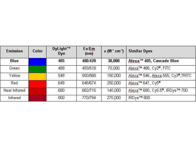
Properties of Dylight(TM) Conjugates.
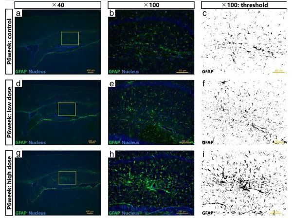
Maternal exposure to Printex 90 carbon nanoparticles dose-dependently increases expression levels of glial fibrillary acidic protein (GFAP) in astrocytes in the offspring hippocampus.a,b,d,e,gandhshow representative fluorescent micrographs of GFAP-positive astrocytes in the hippocampi of 6week old male offspring in the control (a,b), low exposure (d,e), and high exposure group (g,h).a,dandggives an overview of the hippocampus, withb,e, andhproviding enlarged views of panelsa,d, andg, respectively.c,f, andiare grayscale views ofb,e, andh, respectively, for quantification of GFAP expression. Fig. 5. PMID: 30201004.
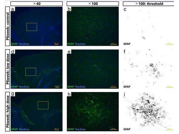
Maternal exposure to Printex 90 carbon nanoparticles dose-dependently increases expression levels of glial fibrillary acidic protein (GFAP) in astrocytes in the offspring cerebral cortex.a,b,d,e,gandhshow representative fluorescent micrographs of GFAP-positive astrocytes in the cerebral cortices of 6week old male offspring in the control (a,b), low exposure (d,e), and high exposure groups (g,h).a,dandggives an overview of the cerebral cortex, withb,e, andhproviding enlarged views hereof, respectively.c,f, andiare grayscale views ofb,e, andh, respectively, for quantification of the GFAP expression. Fig 4. PMID: 30201004.
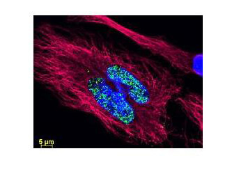
DyLight(TM) dyes can be used for multi-color immunofluorescence microscopy with uniform fluorescence intensity throughout the image. DyLight(TM) dyes are exceptionally bright and photostable and are optimized for microscopy and microarray detection methods. This image shows anti-histone detection using a DyLight(TM) 488 conjugate (green). Anti-Tubulin was detected using a DyLight(TM) 549 conjugate (red). Nuclei were counter-stained using DAPI (blue). The image was captured using an Axio Imager.Z1 (Zeiss Micro Imaging Inc).
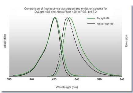
DyLight(TM) 488 Fluorescence Spectra.
* VAT and and shipping costs not included. Errors and price changes excepted