0.02 M Potassium Phosphate, 0.15 M Sodium Chloride, pH 7.2
Form:
Lyophilized
Target:
Goat
Antibody Type:
Secondary Antibody
Application Dilute:
FLISA: >1:20,000, IF Microscopy: >1:5,000, WB: >1:10,000
Application Notes:
The emission spectra for this DyLight(TM) conjugate match the principle output wavelengths of most common fluorescence instrumentation. This product is designed for immunofluorescence microscopy, fluorescence based plate assays (FLISA) and fluorescent weste
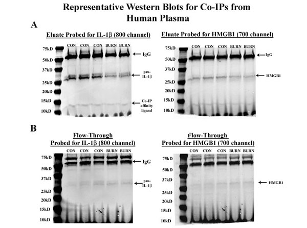
Entire western blots for co-immunoprecipitation of HMGB1 and IL-1beta in human plasma.Co-immunoprecipitation was performed for HMGB1 and IL-1beta in human control and burn plasma. Entire eluate and flow through for these samples are presented. (A) Entire blots of eluates probed for IL-1beta and for HMGB1. Bands positive for IgG and pro-IL-1beta as well as the Catch and Release affinity ligand were observed. The blot probed for HMGB1 showed a broad band consistent with free HMGB1 near 29kD. (B) Entire western blots of flow through. Minimal pro-IL-1beta and HMGB1 were found in the flow through. Fig S4. PMID:29601597.
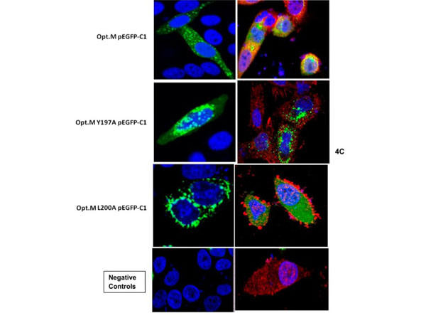
Immunofluorescence results using Donkey Anti-Goat IgG DyLight 800.Effect of mutation in YXXL domain of HRSV M protein.C) The mutation of the tyrosine at amino acid residue 197 to alanine and leucine at amino acid residue 200 to alanine causes a distinct phenotypic change in the localization of the HRSV Matrix protein. HEp2 cells were transfected with indicated pEGFP-C1 Opt. M constructs and were fixed with paraformaldehyde at 24 hours post transfection as described previously. Cells were imaged for Opt.M, adaptor protein and DAPI using CLSM protocols described previously. Negative controls include transfection of cells with empty plasmid vector (pEGFP-C1) without M insert (left side panel) and mock transfected cells with staining for adaptor protein only with goat anti Ap3u3A followed by rabbit anti goat Alexa Fluor 546 antibody (right side panel). The first 3 images on the left side panel shows staining for Opt.M and DAPI. The first 3 images on the right side panel shows merged image with staining for Opt.M, adaptor protein and DAPI. The results were reproducible in at least two independent assays.Fig 4. PMID: 29028839.
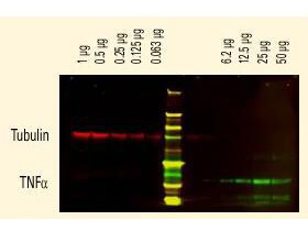
DyLight(TM) dyes can be used for two-color Western Blot detection with low background and high signal. Anti-tubulin was detected using a DyLight(TM) 680 conjugate. Anti-TNFa was detected using a DyLight(TM) 800 conjugate. The image was captured using the Odyssey Infrared Imaging System developed by LI-COR.
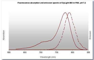
DyLight(TM) 800 Fluorescence Spectra.
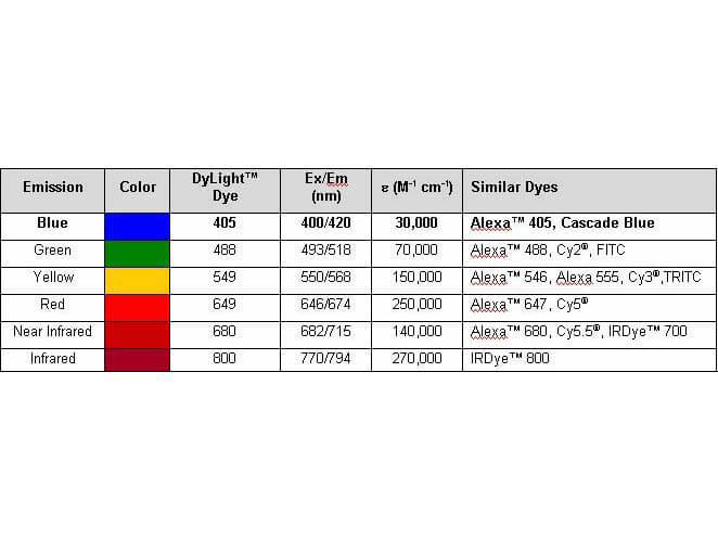
Properties of DyLight(TM) Conjugates.
* VAT and and shipping costs not included. Errors and price changes excepted