0.02 M Potassium Phosphate, 0.15 M Sodium Chloride, pH 7.2
Form:
Lyophilized
Target:
Mouse
Antibody Type:
Secondary Antibody
Application Dilute:
FLISA: >1:20,000, IF Microscopy: >1:5,000, WB: >1:10,000
Application Notes:
This product is designed for immunofluorescence microscopy, fluorescence based plate assays (FLISA) and fluorescent western blotting. This product is also suitable for multiplex analysis, including multicolor imaging, utilizing various commercial platfor
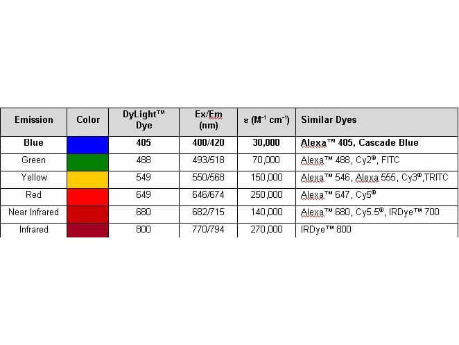
Properties of DyLight(TM) Fluorescent Dyes.
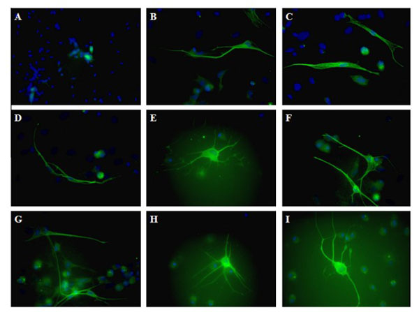
Differentiated rMSCs immunofluorescence. Cells were stained with an antibody against b-III-tubulin (green) using Rabbit Anti-Mouse IgG DyLight(TM)488 (p/n 610-441-002). Differences in the number of primary dendrites and branching dendrites using various differentiation schemes. Negative control (A), control, differentiation using a basic differentiation medium (B and C), long-term imipramine treatment (D), CACM treatment (E), CACM + desipramine (F and G), CACM + fluoxetine (H, I). In each experiment, the nuclei were counterstained with 40 ,6- diamidino-2-phenylindole (DAPI) (blue). In our study, only cells that were positive stained and had the shape that is characteristic for neuronal cells were qualified as positive. (For interpretation of the references to color in this figure legend, the reader is referred to the web version of this article.) Fig. 4. .PMID: 25712637.
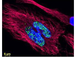
DyLight(TM) dyes can be used for multi-color immunofluorescence microscopy with uniform fluorescence intensity throughout the image. DyLight(TM) dyes are exceptionally bright and photostable and are optimized for microscopy and microarray detection methods. This image shows anti-histone detection using a DyLight(TM) 488 conjugate (green). Anti-Tubulin was detected using a DyLight(TM) 549 conjugate (red). Nuclei were counter-stained using DAPI (blue). The image was captured using an Axio Imager.Z1 (Zeiss Micro Imaging Inc).
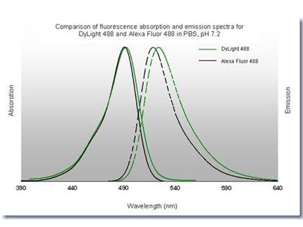
DyLight(TM) 488 Fluorescence Spectra.
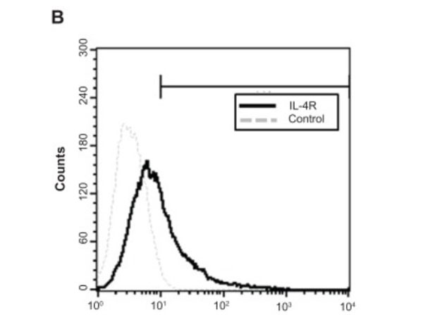
(B) Flow cytometric detection of the cell-surface interleukin-4 receptor on cloned human GBM8401-luc cells. Fig 1. PMID: 22393293.
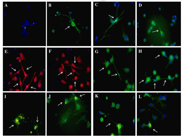
Differentiated rMSCs immunofluorescence. Negative control (A). Cells were stained with an antibody against b-III-tubulin (green) after 14 days (B) and 28 days (C) of differentiation using Rabbit Anti-Mouse IgG DyLight(TM)488 (p/n 610-441-002). After 28 days of differentiation rMSCs were positive for Gad67 (green) (D), choline acetyltransferase (Acht) (red) (E, F) and tyrosine hydroxylase (Th) (green) (G, H). Cells positive for Gad67 (green) also co-expressed Cxcr4 (yellow) (I, J), and cells positive for Th (green) co-expressed Cxcr4 (yellow) as well (K, L). In each experiment the nuclei were counterstained with 40 ,6-diamidino-2-phenylindole (DAPI) (blue). In our study only cells which were positive stained and had the shape characteristic for neuronal cells, were qualified as positive for examined markers (white arrows). (For interpretation of the references to color in this figure legend, the reader is referred to the web version of this article.) Fig. 5. PMID: 25712637.
* VAT and and shipping costs not included. Errors and price changes excepted