0.02 M Potassium Phosphate, 0.15 M Sodium Chloride, pH 7.2
Form:
Lyophilized
Target:
Rat
Antibody Type:
Secondary Antibody
Application Dilute:
FLISA: >1:20,000, IF Microscopy: >1:5,000, WB: >1:10,000
Application Notes:
This product is designed for immunofluorescence microscopy, fluorescence based plate assays (FLISA) and fluorescent western blotting. This product is also suitable for multiplex analysis, including multicolor imaging, utilizing various commercial platfor
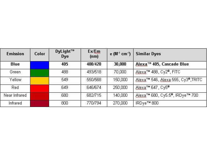
Properties of DyLight(TM) Conjugates.
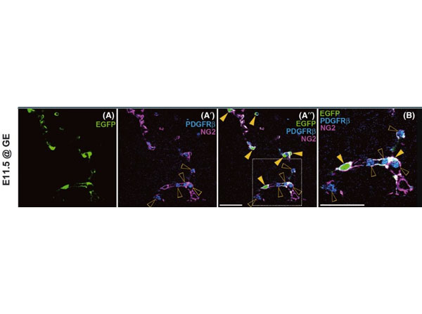
Existence of non-pericyte enhanced green fluorescent protein (EGFP+) cells in the telencephalon of E11.5P0-Cre/EGFPmice. (A-B) A coronal section at the telencephalic level is stained with anti-GFP, PDGFRbeta, and NG2 antibodies. Most of EGFP+cells express both PDGFRbeta and NG2 (yellow arrowheads). (B) High-magnification of the dashed square in A''. PDGFRbeta is also expressed in hematopoietic cells (open arrowheads in A'-B). Fig 5. PMID: 23157329.
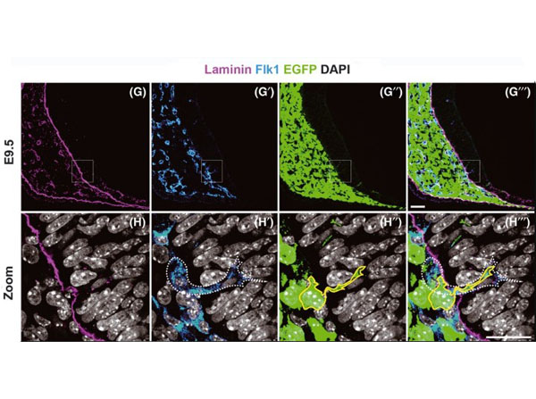
Penetration and distribution of neural crest-derived cells (NCDCs) in theP0-Cre/EGFPmouse telencephalon. (G-H''') A coronal section at the telencephalic level of the E9.5 embryo is stained with anti-laminin, Flk1, GFP antibodies and counter-stained 4', 6-diamidino-2-phenylindole (DAPI). High-magnification images within the squares in G-G''' are shown in H-H'''. Through the laminin+basement membrane (H), an endothelial cell (white dashed line in H') invades the telencephalon together with an EGFP+cell (yellow line in H''). Scale bars: (for G-G'''), 50µm, H''' (for H-H'''), 25µm. Figure 2. PMID: 23157329.
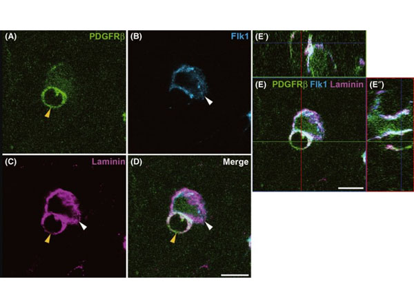
The usability of PDGFRbeta and NG2 as pericyte markers in the telencephalon. (A-E'') Immunostaining with anti-PDGFRbeta, Flk1, and laminin antibodies on coronal section at the telencephalic level of E11.5P0-Cremouse embryo. (A-D) A PDGFRbeta+pericyte (yellow arrowheads) wraps around Flk1+endothelial tube (white arrowheads), and these cell types are surrounded by laminin+basement membrane. This pattern is also clearly observed in orthogonal view of the image shown in D (E-E''). Fig 3. PMID: 23157329.
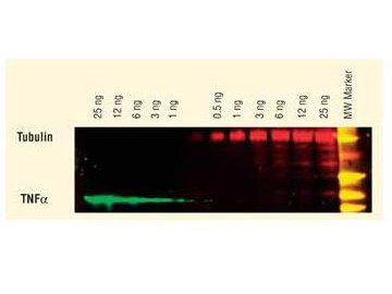
DyLight(TM) dyes can be used for two-color Western Blot detection with low background and high signal. Anti-tubulin was detected using a DyLight(TM) 549 conjugate. Anti-TNFa was detected using a DyLight(TM) 649 conjugate. The image was captured using the Typhoon(TM) 9410 Imaging System.
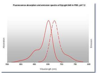
DyLight(TM) 649 Fluorescence Spectra.
* VAT and and shipping costs not included. Errors and price changes excepted