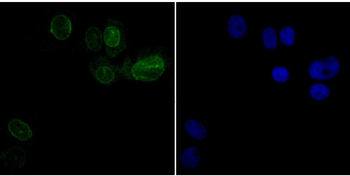
ICC staining SUN2 (green) in SK-Br-3 cells. The nuclear counter stain is DAPI (blue). Cells were fixed in paraformaldehyde, permeabilised with 0.25% Triton X100/PBS.
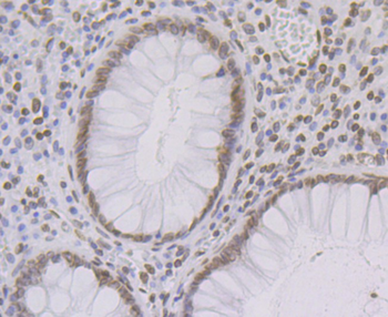
Immunohistochemical analysis of paraffin-embedded human colon tissue using anti-SUN2 antibody. Counter stained with hematoxylin.
Immunohistochemical analysis of paraffin-embedded human prostate cancer tissue using anti-SUN2 antibody. Counter stained with hematoxylin.
Immunohistochemical analysis of paraffin-embedded mouse liver tissue using anti-SUN2 antibody. Counter stained with hematoxylin.
Immunohistochemical analysis of paraffin-embedded rat epididymis tissue using anti-SUN2 antibody. Counter stained with hematoxylin.
Western blot analysis of SUN2 on A431 cell lysates with Rabbit anti-SUN2 antibody (orb1499408) at 1/500 dilution. Lysates/proteins at 10 µg/Lane. Predicted band size: 80 kDa, Observed band size: 75 kDa, Exposure time: 2 minutes, 8% SDS-PAGE gel. Proteins were transferred to a PVDF membrane and blocked with 5% NFDM/TBST for 1 hour at room temperature. The primary antibody (orb1499408) at 1/500 dilution was used in 5% NFDM/TBST at room temperature for 2 hours. Goat Anti-Rabbit IgG - HRP Secondary Antibody at 1:300000 dilution was used for 1 hour at room temperature.
* Mehrwertsteuer und Versandkosten nicht enthalten. Irrtümer und Preisänderungen vorbehalten