Application Notes: 1 µg/ml of SMC-479 was sufficient for detection of TCP1 alpha in 20 µg of 3T3 cell lysate by colorimetric immunoblot analysis using Goat anti-rat IgG:HRP as the secondary antibody
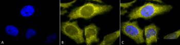
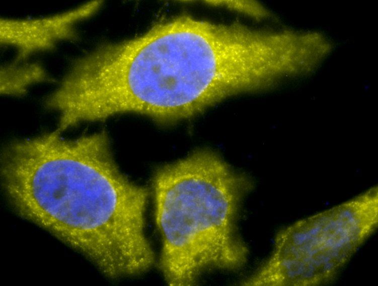
Immunocytochemistry/Immunofluorescence analysis using Rat Anti-TCP1-alpha Monoclonal Antibody, Clone 91a. Tissue: Heat Shocked cervical cancer cells (HeLa). Species: Human. Fixation: 2% Formaldehyde for 20 min at RT. Primary Antibody: Rat Anti-TCP1-alpha Monoclonal Antibody at 1:100 for 12 hours at 4C. Secondary Antibody: R-PE Goat Anti-Rat (yellow) at 1:200 for 2 hours at RT. Counterstain: DAPI (blue) nuclear stain at 1:40000 for 2 hours at RT. Localization: Cytoplasm. Centrosome. Magnification: 100x. (A) DAPI (blue) nuclear stain. (B) Anti-TCP1-alpha Antibody. (C) Composite. Heat Shocked at 42C for 1h.
Western Blot analysis of Human A431 and HEK293 cell lysates showing detection of TCP1 alpha protein using Rat Anti-TCP1 alpha Monoclonal Antibody, Clone 91a. Primary Antibody: Rat Anti-TCP1 alpha Monoclonal Antibody at 1:1000.
Immunocytochemistry/Immunofluorescence analysis using Rat Anti-TCP1-alpha Monoclonal Antibody, Clone 91a. Tissue: Heat Shocked cervical cancer cells (HeLa). Species: Human. Fixation: 2% Formaldehyde for 20 min at RT. Primary Antibody: Rat Anti-TCP1-alpha Monoclonal Antibody at 1:100 for 12 hours at 4C. Secondary Antibody: APC Goat Anti-Rat (red) at 1:200 for 2 hours at RT. Counterstain: DAPI (blue) nuclear stain at 1:40000 for 2 hours at RT. Localization: Cytoplasm. Centrosome. Magnification: 20x. (A) DAPI (blue) nuclear stain. (B) Anti-TCP1-alpha Antibody. (C) Composite. Heat Shocked at 42C for 1h.
Immunofluorescence analysis of heat shocked hela cells using TCP1-alpha antibody
Immunofluorescence analysis of heat shocked hela cells using TCP1-alpha antibody
* Mehrwertsteuer und Versandkosten nicht enthalten. Irrtümer und Preisänderungen vorbehalten