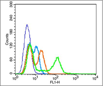
Flow cytometric analysis of EC9706 Cell using AKT1 (phospho-Tyr315-Tyr316-Tyr312) antibody.
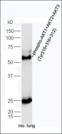
Western blot analysis of mouse lung tissue using AKT1 (phospho-Tyr315-Tyr316-Tyr312) antibody.
Blank control (blue): EC9706 (fixed with 2% paraformaldehyde for 10 min at 37C). Primary Antibody: Rabbit Anti-phospho-AKT1+AKT2+AKT3 (Tyr315+316+312) antibody (orb6789, Green), dilution: 1 µg in 100 µl 1X PBS containing 0.5% BSA, Isotype Control Antibody: Rabbit IgG (orange), used under the same conditions, Secondary Antibody: Goat anti-rabbit IgG-FITC (white blue), dilution: 1:200 in 1X PBS containing 0.5% BSA.
Paraformaldehyde-fixed, paraffin embedded (Mouse brain), Antigen retrieval by boiling in sodium citrate buffer (pH6.0) for 15 min, Block endogenous peroxidase by 3% hydrogen peroxide for 20 minutes, Blocking buffer (normal goat serum) at 37C for 30 min, Antibody incubation with (phospho-AKT1+AKT2+AKT3 (Tyr315+316+312)) Polyclonal Antibody, Unconjugated (orb6789) at 1:500 overnight at 4C, followed by a conjugated secondary for 20 minutes and DAB staining.
Sample: Heart (Mouse) Lysate at 40 ug, Primary: Anti-phospho-AKT1+AKT2+AKT3 (Tyr315+316+312) (orb6789) at 1/300 dilution, Secondary: IRDye800CW Goat Anti-Rabbit IgG at 1/20000 dilution, Predicted band size: 56 kD, Observed band size: 63 kD.
Sample: Lane 1: Adrenal glands (Mouse) Lysate at 40 ug, Lane 2: Cerebrum (Mouse) Lysate at 40 ug, Primary: Anti-phospho-AKT1+AKT2+AKT3 (Tyr315+316+312) (orb6789) at 1/1000 dilution, Secondary: IRDye800CW Goat Anti-Rabbit IgG at 1/20000 dilution, Predicted band size: 60 kD, Observed band size: 60 kD.
Sample: Lane 1: Rat Cerebrum tissue lysates, Lane 2: Rat Heart tissue lysates, Primary: Anti-phospho-AKT1+AKT2+AKT3 (Tyr315+316+312) (orb6789) at 1/1000 dilution, Secondary: IRDye800CW Goat Anti-Rabbit IgG at 1/20000 dilution, Predicted band size: 56 kDa, Observed band size: 65 kDa.
* Mehrwertsteuer und Versandkosten nicht enthalten. Irrtümer und Preisänderungen vorbehalten