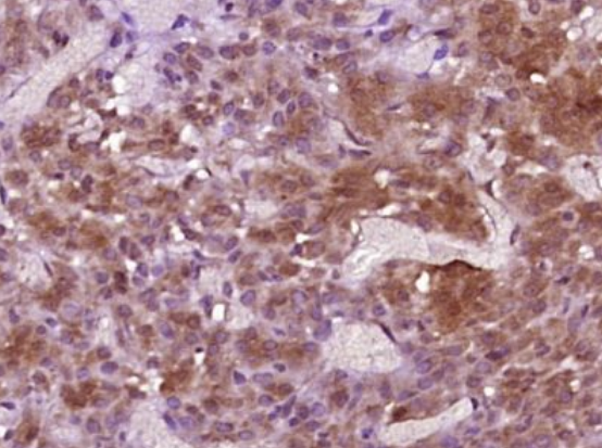
Immunohistochemical analysis of rat ovary tissue using Inhibin beta B antibody
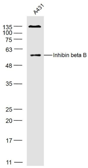
Western blot analysis of A431(Human) Cell Lysate using Inhibin beta B antibody.
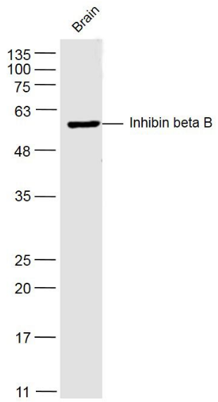
Western blot analysis of Brain (Mouse) Lysate using Inhibin beta B antibody.
Paraformaldehyde-fixed, paraffin embedded (rat ovary tissue), Antigen retrieval by boiling in sodium citrate buffer (pH6.0) for 15 min, Block endogenous peroxidase by 3% hydrogen peroxide for 20 minutes, Blocking buffer (normal goat serum) at 37C for 30 min, Antibody incubation with (Inhibin beta B) Polyclonal Antibody, Unconjugated (orb13510) at 1:400 overnight at 4C, followed by operating according to SP Kit (Rabbit) instructionsand DAB staining.
Sample: A431 (Human) Cell Lysate at 40 ug, Primary: Anti-Inhibin beta B (orb13510) at 1/300 dilution, Secondary: IRDye800CW Goat Anti-Rabbit IgG at 1/20000 dilution, Predicted band size: 45 kD, Observed band size: 50 kD.
Sample: Brain (Mouse) Lysate at 40 ug, Primary: Anti-Inhibin beta B (orb13510) at 1/300 dilution, Secondary: IRDye800CW Goat Anti-Rabbit IgG at 1/20000 dilution, Predicted band size: 45 kD, Observed band size: 50 kD.
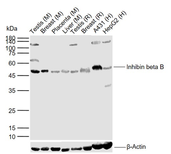
Sample: Lane 1: Mouse Testis tissue lysates, Lane 2: Mouse Breast tissue lysates, Lane 3: Mouse Placenta tissue lysates, Lane 4: Mouse Liver tissue lysates, Lane 5: Rat Testis tissue lysates, Lane 6: Rat Breast tissue lysates, Lane 7: Human A431 cell lysates, Lane 8: Human HepG2 cell lysates, Primary: Anti-Inhibin beta B (orb13510) at 1/1000 dilution, Secondary: IRDye800CW Goat Anti-Rabbit IgG at 1/20000 dilution, Predicted band size: 13/45 kDa, Observed band size: 46 kDa.
* VAT and and shipping costs not included. Errors and price changes excepted